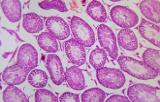
Routine tissue stains
We distinguish between routine stains that are performed on almost all samples for a first study and specific stains that are performed to obtain more information when routine staining is not enough.
Hematoxylin and Eosin (H & E) staining is routinely used in histopathology laboratories as it allows pathologists and researchers to obtain a very detailed view of the tissue. This is because staining of cell structures such as cytoplasm, nucleus, organelles and extracellular components is performed. This information is sometimes sufficient to establish a diagnosis of the organization (or disorganization) of the cells of the tissue and also shows the abnormalities or the particular indicators of the cells (like the changes of the nucleus typical of certain cancers). Even when other more complex methods of staining are used, H & E staining continues to be part of the diagnosis because it allows one to see the morphology of the tissue which will then allow the pathologist or researcher to interpret the other stains.
In the laboratories, all the samples are initially stained by the H & E method and the specific stains are carried out only if it is necessary to have more information to obtain a more detailed analysis, for example to differentiate 2 types of cancer having a similar morphology.


