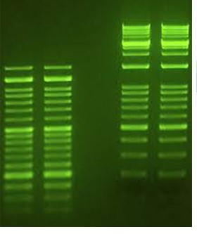
Neodye DNA Green (35,000 ×)
Catalogue number NB-60-0009
Presentation 1 mL
|
Description Neodye DNA Green (35,000 ×) presents a safer alternative to ethidium bromide for detecting nucleic acids in agarose gels. With comparable sensitivity, it seamlessly integrates into agarose gel electrophoresis protocols, emitting green fluorescence upon binding to DNA or RNA. This advanced stain showcases two secondary fluorescence excitation peaks at approximately 270 nm and 290 nm, alongside a robust excitation peak at 490 nm. Its fluorescence emission closely resembles that of ethidium bromide when bound to DNA, peaking at around 530 nm, ensuring compatibility with a wide range of gel reading instruments. Embrace the next generation of nucleic acid staining with Neodye DNA Green (35,000 ×) - where safety,sensitivity, and compatibility converge. |
 |
Features
▪ Detects double-strand DNA and single-stranded RNA effectively.
▪ Offers a safe alternative to ethidium bromide staining.
▪ Exhibitssensitivity on par with EtBr.
▪ Non-toxic, non-mutagenic, and non-carcinogenic composition.
▪ Produces no hazardous waste, ensuring environmental friendliness.
Shipping & Storage Conditions
This product can be shipped from Blue Ice to Room temperature. Upon receipt, store Neodye DNA Green (35,000 ×) at Room temperature or at
2°C to 8°C protected from light. Storage at temperatures below may degrade Neodye DNA Green (35,000 ×).
| COMPONENT | TUBES | VOLUME |
| Neodye DNA Green (35,000 ×) | 1 | 1 mL |
Standard Protocol
Pre-staining protocol
1. Prepare 70 -100 mL of an agarose gel solution (concentration from 0.8-3.0%) and heat until the solution is completely clear, and no small
floating particles are visible.
2. Let the solution cool down and add 2-3 µL of Neodye DNA Green (35,000 ×) to the gelsolution.
3. Mix gently and cast into the tray.
4. When the gel issolid, load the samples and perform electrophoresis.
5. Detect the bands under anUV trans-illuminator.
Post-staining protocol
1. For <0.5 cm thick agarose gels, add 10-15 μL ofstain per 100 mL of buffer. Please notice that the amount ofstain may depend on the thickness
of the gel and the percentage of agarose.
2. Staining time can range from 5 to 60 minutes.
3. The post-staining solutionmay be used 2-3 times. Staining solution to be reused should preferably be stored at room temperature in the dark.

